Tuesday, February 28, 2012

If you give a mouse a placebo...

...It might ask for some cocaine.  Or it might feel the effects of cocaine anyway. 
Just say no, Rat (source)

The "Placebo Effect" occurs when someone takes a functionally ineffectual drug, but feels the effects anyway. There are many examples of this: Someone in pain takes a sugar pill, but is told that it is a painkiller might report 'feeling much less pain'.  A Parkinson's patient takes a sugar pill having been told it was their 'L-dopa' medication and can suddenly move more fluidly. The "Placebo Effect" is so strong that most experiments testing the effectiveness of a drug in healing anything use a placebo control.  The researchers want to make sure that the drug has an actual effect that is greater than the placebo effect.  (It has been proposed that homeopathic remedies are entirely due to the placebo effect)

One problem with deeply understanding the physical mechanisms which underlie the placebo effect is that all the experiments must be on humans.  You can't simply tell a mouse it's getting a 'cure' and give it a fake pill.  However, scientists at the National Institute on Drug Abuse (NIDA) have conducted an ingenious experiment that involves giving a mouse what is essentially a placebo.  Better still, they published it in PLoS One, so everyone can read the paper for free!

Before we dive into the placebo aspect of this paper, we need to back up and learn a little about as the addiction and reward system works in the brain.

In 1954, James Olds and Peter Milner published a paper showing that a rat would press a lever to receive an electrical stimulation in certain areas of its brain.
Olds and Milner, 1954 Figure 2, Xray of rat
This was a huge discovery showing that 'reward' could be activated directly. 

Later studies found that when this electrode stimulates the dopamine system of a rat, the rat will press and press and press this lever, even forgoing food when it is famished.  Incidentally, a mouse/rat will also compulsively press a lever to get injections of cocaine (which acts by stimulating the dopamine system).  You can do all sorts of experiments on drug addiction using this cocaine self-injection system. You can test how long it takes the mouse to become addicted, you can test the effect of drug concentration, you can test how other drugs interact with self-injection of cocaine, and you can even test aspects of withdrawal and relapse.

Which brings up back to our placebo.  Wise et al., (2008) investigated the mechanisms underlying this self-administration.  What was happening in the mouse brain when they got a dose of cocaine? They found that when the mouse pressed the lever and got the cocaine there was a surge of dopamine almost immediately after.  There is a center in the brain called the VTA that contains neurons which release dopamine. When these neurons are active, other areas of the brain are flushed with dopamine and the person/rat/mouse 'feels reward'.  But what makes these neurons active?

This brings up a problem we discussed a while ago, about the never ending cycle of neuronal firing.  The dopamine neurons fire, but why? what neurons are firing onto them to make them fire, and then, what neurons are making those neurons fire and which ones are firing before that...so forth into forever. 
To go one step up in this firing-chain, Wise et al. cleverly looked at 'brain juice' in the VTA and found that when the cocaine is administered, the mouse gets a surge of glutamate there.  (Glutamate activates cells, so this would cause the dopamine neurons of the VTA to fire and release dopamine onto other cells). 

So what does all this mean, and how does it get us to a placebo for a rat?

Here's the thing: the surge of glutamate that stimulates the VTA only shows up in mice that have already learned that a lever press gives them cocaine.  (That is, this glutamate surge doesn't occur the very first time the mouse gets cocaine)

Wise et al., 2008 (figure1B)
 Here is the figure showing this glutamate surge in the VTA.  The vertical dotted gray line is when the mouse presses the lever for the cocaine.  The red and yellow traces are the condition where the mouse actually gets cocaine in response to the lever press.  the blue and green traces are the condition where the mouse gets saline instead (a control), and the gray trace is the first time the rat gets cocaine in response to the lever press.

 Another peculiar aspect of this glutamate surge is that it is probably too fast a response to be a result of the cocaine acting in the brain.  So how is the injection of cocaine causing a glutamate surge if it is not even acting on the brain yet?  This is quite the puzzle.  Wise et al., wanted to make sure that this glutamate surge was absolutely not due to the cocaine reaching the brain, so they invented the rat placebo! They altered the cocaine molecule, so it was mostly the same shape as normal cocaine, except it couldn't cross the blood brain barrier.  That is, when it is injected into the mouse, it can be detected in the blood and in the peripheral body, but it won't be detected in the brain, and the cocaine will not be able to act directly on the neurons.

When they run the test with this molecule injected instead of cocaine, the figure looks like this:

Wise et al., 2008 (figure1E)
Pretty similar! This cocaine molecule can't reach the brain, but it causes the same glutamate surge as the real stuff! This shows that glutamate surge is somehow due to the cocaine being felt by the body, but not being felt by the brain.  Although they don't call it a placebo in the paper, that is essentially what it is.  It is tricking the brain into thinking it has just received cocaine. (In a stronger way than context can, as evidenced by the lack of response to saline.) 

 So there you have it, a way to trick a rat into thinking it has just received a 'real drug' when it has actually received an ineffectual drug. I think this technique could be adapted to actually study the physiological mechanisms governing the placebo effect. 

© TheCellularScale

ResearchBlogging.orgWise RA, Wang B, & You ZB (2008). Cocaine serves as a peripheral interoceptive conditioned stimulus for central glutamate and dopamine release. PloS one, 3 (8) PMID: 18682722

Thursday, February 23, 2012

Know thyself, Cell: Neuronal self-recognition

A neuron's shape is important for its function, but how does it get its shape in the first place? As we've discussed before, dendrites grow out of the cell body (soma) and follow a somewhat pre-described pattern.  A Purkinje cell always has a general corral-like shape, but each individual neuron is shaped a little differently.  (Just like an oak tree looks different from a pine tree, while at the same time no two oak trees are exactly the same.) 
branching tree at sunset, San Diego: taken by me

And just as trees branch and grow based on where the sunlight is coming from, dendrites can branch and grow depending on external factors.  Of course dendrites don't care about sunlight, but they do want to efficiently 'cover space' to receive lots of incoming signals. 

So how do they do it?

Some neurons have dendrites that repulse eachother. As in, if two dendrites are rooted to the same soma, those two dendrites will avoid eachother.  This is one way that the dendrite can 'cover space' very efficiently, it will branch and grow until it sees 'itself' and then it will stop and grow in a different direction. 

(source) The Leech: yuck.


In 1998, Wang and Macagno published a fascinating study using the mechanosensory neurons in the leech. These particular neurons show 'self-avoidance' (the dendrites of the same neuron do not overlap), but they don't show 'class-avoidance' (Dendrites from the same class of neurons do overlap).
Wang and Macagno wanted to test what exactly was so self-repulsive about these dendrites. 

There are two ways the cell could recognize itself:

1. Through the use of external signals (such as a chemical marker on the surface of the dendrites that sibling dendrites can detect)
2. Through the use of internal signals (such as the voltage activity transmitted within the cell)

To find out what method the dendrites are using to recognize themselves, Wang and Macagno used a laser to separate a small section of dendrite from the rest of the neuron.  Would the other dendrites still avoid this severed dendrite, or would they suddenly see it as a stranger and start to overlap with it?

Wang and Macagno, 1998 Figure 4

The attached dendrites start to treat the severed dendrite as a stranger, growing into its area and overlapping with it.  Figure 4 from Wang and Macagno shows the intact cell (A), the location of the cut (star in B), and the regrowth of both the severed dendrite and the still attached dendrite (arrow in D).

The authors offer several possible explanations for why a severed dendrite would appear to be a stranger to the rest of the dendrites, but all are speculative.  Maybe the electrical signal prevents gap junctions from forming. Maybe there are channels (such as NMDA receptors) that repulse each other when they are both active at the same time.  Maybe there is some cytoplasmic molecule that diffuses between the dendrites and prevents overlap (though the authors admit this mechanism sound too slow to do the job.)  The authors even find a problem with the electrical signal hypothesis in that:
"In some systems the blockade of electrical activity does not affect morphogenesis."

Since no one has tested a blockade of electrical activity on these neurons, the mechanism underlying the self-repulsive nature of these dendrites is still a mystery.
© TheCellularScale

ResearchBlogging.org
Wang H, & Macagno ER (1998). A detached branch stops being recognized as self by other branches of a neuron. Journal of neurobiology, 35 (1), 53-64 PMID: 9552166

Sunday, February 19, 2012

Neurosexism and Delusions of Gender

On the cellular scale, it is very difficult, if not impossible, to tell the brains of men and women apart.  That is, if you zoom in on a part of the brain (like the hippocampus, cortex or striatum) and look at the morphology of a single neuron or the electrical characteristics of that neuron, you would be hard pressed to tell if the neuron you are looking at belongs to a male or a female. This is not very surprising since it is also difficult to tell if the neuron you are looking at is from a human, ape, or elephant

That is a cellular introduction to the non-cellular book that I am reviewing today: Delusions of Gender: How our minds, society, and neurosexism create difference by Cordelia Fine. 


My friend from undergrad who is now a philosophy professor recommend this book to me and described it in such a way that I suspected I would hate it. Upon actually reading it however, I was quite surprised at how informative and entertaining it was. 

 Let's get to it!
Delusions of Gender is a book written in response to the idea that there are inherent differences between men and women that are hard-wired into the brain by evolution and that make women naturally suited for certain activities and men naturally suited for others. 

In other words, Cordelia Fine claims that this is NOT the case, or at least that it is not nearly as much the case as people currently believe.  This is a difficult claim to make.  It's so easy to see that men and women are different in body, so why wouldn't their brains be different? And doesn't evolution make the sexes of many species 'inherently' different? For examples, look at the praying mantis, the zebra finch, and the stickleback. 

So what I found amazing is that Cordelia Fine argued this impossible claim so well that I was thoroughly convinced that everything I had heard about the differences between male and female brains and abilities was at best uncertain, and at worst completely wrong. 

Let me summarize some of the exact points that she makes:

  1. You are bound to find differences when you are looking for them.
  2. Differences are more likely to be reported and publicized than similarities.
  3. There are glaring flaws in many neuroscience studies showing brain differences between men and women.
  4. Even if all the studies showing brain differences between men and women were taken as true, that still wouldn't mean that the differences are 'hard wired' or 'inherent' or 'because of evolution'
  5. Even if all the brain differences are real, and even if they are 'hard wired', that still doesn't mean that women and men actually think differently. 

To explain a little further:

Claim 1 says that when there is one difference between two groups, it is easy to think of a scientific study to determine if something else is different between these two groups.  This is important because of the 'obvious' differences between men and women (yeah, they have different junk). Since everyone knows men and women are different re: genitalia, let's test whether they are different in brain or behavior.  This may seem totally reasonable, but a counter example is finger-print pattern.  People can be grouped by their fingerprint pattern into 'loop-shape' or 'swirl-shape' people.  This fingerprint pattern is determined genetically, but since it is not an obvious difference (you probably don't even know which group you belong to), no one has ever tested whether 'loop-shape' people have bigger hippocampi than 'swirl-shape' people. 

Claim 2 says that for every newspaper headline shouting "Women are inherently totally whacked"  (see also this post) there might be several studies quietly showing "Women and men are pretty similar" This problem, research showing differences getting published and hyped, while research showing 'no difference' getting ignored or not even published to begin with is not a problem new to science, nor is it specific to gender issues. It is always more exciting and always has a 'higher impact' as they say, to show that two things are different. 

A recent paper by De Liberto et al., (2012) shows that male and female mice show no difference in the amount of DAT (dopamine transporter) in the striatum

De Liberto et al., 2012 figure1C
Of course this finding is not the basis for the paper, and it could never be because no journal would publish something so boring.  The point of the paper is that when you apply an estrogen-like substance, you do see a gender difference in how these cells react.

Let's do a quick thought experiment:  Imagine that the researchers had found a difference here, say "male mice have less DAT than female mice."  This could have 'meant' that men have more dopamine at the synapse (as the DAT is responsible for cleaning up excess dopamine there), and this could have been 'translated' into the idea that men are happier or women are more moody and prone to depression (Dopamine levels are implicated in moodiness and depression).  Wow! what an exciting finding!  Front cover of Time magazine: "The Secret Science Behind Moody Women." However, here in reality, these results showed no difference, so they became just a small figure panel in paper about the effects of estrogen. 

Claim 3 is the one I had the hardest time reading.  It is true that there is a lot of sloppy science out there, but I think she goes a little too far in her distrust of science.  For example, she brings up the dead fish in an MRI study as evidence for fMRI studies being flawed.  It is true that in all scientific studies there is a risk of falsely identifying a difference when there is none, but that is exactly what the correction for multiple comparisons is for.  This is a statistical correction that all scientist should know about and apply, but sometimes (maybe even often) they don't.  However, there are plenty of fMRI studies that do correct for this multiple testing problem and are scientifically sound.

Claims 4 and 5 are my favorite.  I thought I would hate this book when I thought that her claim was going to be 'men and women are not different, and neuroscience is flawed and stupid.'  However, when I saw that she was actually claiming 'we don't know enough about the brain to draw the conclusions people are drawing' I got right on board.  There is way too much "the amygdala lit up therefore the person was frightened" and "the hippocampus is bigger so the person must navigate space better" going around. These claims can get ridiculous and most are just not supported.  We don't have a full understanding of the brain, or even of any part of the brain.  We don't even have a full understanding of the single neurons that make up the brain (as you well know from reading The Cellular Scale).

In conclusion, there may be (and probably are) brain differences between men and women, some of these differences might be 'hard wired' and 'inherent' and some of them might develop as a child grows up in a gender-difference driven culture. (By the time you get a kid into an MRI machine, s/he has done a lot of developing.) These (possible/probable) brain differences might mean that men and women think differently, or they might not.
We just don't know enough about it, yet.

© TheCellularScale
There are many excellent reviews of this book out there, here are just a few:
SevenDeadlySynapses
NeuroSkeptic (who is actually cited in this book)
TheThinkingMeatProject
GenderAccrossBorders


ResearchBlogging.orgDi Liberto V, Mäkelä J, Korhonen L, Olivieri M, Tselykh T, Mälkiä A, Do Thi H, Belluardo N, Lindholm D, & Mudò G (2012). Involvement of estrogen receptors in the resveratrol-mediated increase in dopamine transporter in human dopaminergic neurons and in striatum of female mice. Neuropharmacology, 62 (2), 1011-8 PMID: 22041555


Thursday, February 16, 2012

Sound localization: form meets function in the BirdBrain

In the last post,  we introduced the special football-shaped cells of the bird Nucleus Laminaris (NL).
Today we look at how the dendritic length of these neurons dictates the frequencies they are most sensitive to.  But first we need to understand what the NL does. 
(source)

When you hear a noise, you can tell what direction it is coming from (for the most part).

There are several ways the brain can hone in on the direction of a sound. One of those ways is called the 'inter-aural time difference.' That means the difference between when a sound hits one ear and when it hits the other.
For example, imagine you are on a hike in a quiet woods and you hear a leaf-rustling sound to your right. The sound waves from that leaf-rustling actually reach your right ear a fraction of a second before they reach your left ear. The brain can process this time difference and compute the direction of the sound. 
 
Incidentally, birds like the boring-old-chicken and the pretty-cool-barn-owl are particularly good at sound localization. The NL in these birds is where this computation takes place.

Figure 1a, Wang and Rubel (2008)

Above is a diagram of the bird auditory brain stem.  the NL is the line-like structure on either side.  The dendrites on the top of the NL neurons receive input from one ear (the ear on the same side of the head), and the dendrites on the bottom of the NL receive input from the other ear (the one on the opposite side of the head).  In the picture above, inputs from one ear are in black and inputs from the other are in red. Since a sound from a particular direction hits one ear first, and then the other ear, the top dendrites of a cell will receive inputs at a slightly different time than the bottom dendrites of that cell.  
Figure 2a, Wang and Rubel (2008)

If you remember our last post, you know that the dendrites in the NL are longer near the outside edges of the brain and shorter near the middle.  You also will remember that the short-dendrite neurons are sensitive to high frequency sound waves and the long-dendrite neurons are sensitive to lower frequency sound waves. So how do the frequency-tuning properties relate to the dendritic gradient properties?

In a computational study, published in Biological Cybernetics, Grau-Serrat et al., (2003) try to answer this question. 

First a quick introduction to computational neuroscience (I plan to write a whole post about this someday):
"Computational neuroscience is a discipline that aims to understand how information is processed in the nervous system by developing formal models at many different structural scales...The end product of an computational analysis should be a sufficiently specified model, internally consistent and complete enough to enable formal mathematical characterization or computer simulation."  Grau-Serrat et. al., 2003
I thought these passages from their introduction made an excellent summary of computational neuroscience. 

Now back to the BirdBrain!

So to answer the 'form and function' question inherent in the NL cells, Grau-Serrat et al. create model NL neurons in a computer program and test them with simulated inputs.  Because it is a computational model, they have complete control over the conditions.  They can put either short or long dendrites on these cells, and can send in high or low frequency sound waves.  They also can send an input to the top layer of dendrites at one time and then send an input to the bottom layer of dendrites milliseconds afterwards.  Controlling all these factors, they can test whether the size of the dendrites in these cells helps them accurately detect the timing difference between ears. They can also test whether the frequency of the sound (high or low) has anything to do with timing difference detection.

They found a remarkably simple answer:
The lower the frequency, the longer the dendrites need to be to show good time discrimination.

In their computational model, making the dendrites longer generally improved time discrimination, but for every frequency there was a certain dendritic length that was 'long enough'.  Adding more dendrite after a certain length didn't improve the time discrimination.

Figure 3, Grau-Serrat et al., 2003

In summary, the dendritic gradient of the NL is predicted if the system follws two rules:
  1. Keep the dendrites as short as possible. 
  2. Make the dendrites long enough to accurately discriminate time differences.

So there you have it, the mystery of the NL dendritic gradient, solved by computational neuroscience.

Note: I know this is a pretty complex system, and I am definitely simplifying. If you know a lot about the auditory brainstem, please don't hesitate to correct/expand on what I've written here in a comment.  Also if you don't know a lot about this system and have a question, write it in the comments section and I'll try my best to answer it.

© TheCellularScale
  
ResearchBlogging.org Grau-Serrat V, Carr CE, & Simon JZ (2003). Modeling coincidence detection in nucleus laminaris. Biological cybernetics, 89 (5), 388-96 PMID: 14669019


ResearchBlogging.orgWang Y, & Rubel EW (2008). Rapid regulation of microtubule-associated protein 2 in dendrites of nucleus laminaris of the chick following deprivation of afferent activity. Neuroscience, 154 (1), 381-9 PMID: 18440716

Monday, February 13, 2012

Neurons tuned like the strings of a harp

The auditory brainstem of the boring-old-chicken is actually home to some fascinating neurons.

Key West rooster, taken by me.

The Nucleus Laminaris (NL) is a group of coincidence-detecting neurons which receive indirect input from both ears and is located in the bird auditory brainstem.

NL neurons show a peculiar dendrite pattern.  These bipolar neurons fall into the particular category of football shaped cells which have dendrites coming out the top and bottom of their cell body. The cell body (soma) of these neurons are about the same size, but depending on where they are in the NL, the cells have either short, medium or long dendrites. 

The ones near the midline have a bunch of short stubby little dendrites.
Figure 2B from Smith and Rubel, 1979

If they are a little further out from the midline, they have longer dendrites.
Figure 3B from Smith and Rubel, 1979
and finally if they are furthest from the middle, they have fewer and much longer dendrites.
Figure 10A Smith and Rubel 1979
all together this makes a gradient from short to long dendrites.  
From Figure6 Smith and Rubel 1979
The big question here is "Why?"

What is the purpose of having stubby or extended dendrites like this?  Well, even in 1979 when Smith and Rubel reconstructed these neurons, they knew that these neurons had a special answer to the "form and function" question.


The amazing thing about these neurons is that they are 'tuned' to respond maximally to specific frequencies (sound waves).  And just like strings on an instrument, the cells with shorter dendrites respond to higher frequencies and the cells with longer dendrites respond to lower frequencies. 

Why is this? Dendrites don't actually vibrate like strings, but there must be some reason for a cell with short dendrites to respond to higher frquencies and a cell with long dendrites to respond to low frequencies. 

The answer lies in what the Nucleus Laminaris actually does. In the next post we'll venture into the wilds of computational neuroscience and explore the reason behind this strange connection between dendrite shape and cell function. 


ResearchBlogging.orgSmith DJ, & Rubel EW (1979). Organization and development of brain stem auditory nuclei of the chicken: dendritic gradients in nucleus laminaris. The Journal of comparative neurology, 186 (2), 213-39 PMID: 447882

Thursday, February 9, 2012

LTP and LTD at the same time? Adventures in Functional Compartmentalization

On Monday we talked about LTP and LTD on a basic level, today we are discussing how they interact with each other.  In a recent Open Access paper, Pavlowsky and Alarcon ask the question: Can some synapses on a neuron strengthen while at the same time others weaken?  And if so, how do the two processes interact with each other?

neurons firing (source)


First let's get some background.  Synapse strengthening (LTP) and synapse weakening (LTD) both require new proteins to be synthesized at the soma* (*in this particular situation, sometimes they don't require it, but those details are too deep to dive into here).  So what happens if LTP is induced at some synapses and LTD is induced at others on the same neuron?  There are three possibilities:

  1. They compete for protein synthesis at the soma, one using up all the precious protein synthesis machinery and impairing the development of the other
  2. They cooperate, one starting up the protein synthesis engine at the soma so it's ready to go, helping the other.
  3. They don't interact and just do their own thing like normal.

To determine which of these possibilities actually happen in a neuron, Pavlowsky and Alarcon induce LTP and LTD on the same cells, but in different places. 

Showing stimulation on the same side of the soma
From Figure 2 Pavlowsky and Alarcon 2012


 They induced LTP in one spot (S2) and then induced LTD in another (S1).  And lo and behold! the LTP happening first prevented the LTD (favoring the compete hypothesis above).

So this shows that LTP and LTD compete in the neuron.  But what are they competing for?

There are two steps in protein synthesis where LTP and LTD might compete: translation (getting a protein from an mRNA) and transcription (creating more mRNA from the DNA). 

To test whether translation or transcription is important for this competition, the researchers induced LTP at S2 in the presence of either a translation blocker (anisomycin) or a transcription blocker (actinomycin-D).  Then they washed away the blocker and induced LTD at S2.


From Figure 6 Pavlowsky and Alarcon 2012


 The translation blocker allowed for subsequent LTD at S1 (top left of figure), while the transcription blocker didn't (top right of figure), even though both prevented the initial LTP at S2 (bottom panels of figure).  This is evidence that the translation phase of  protein synthesis is important for determining which form of plasticity gets induced (LTP or LTD). 

So what does all this mean? The results support the compete hypothesis, that the first plasticity induction (LTP or LTD) gets dibs on most of the plasticity-related protein synthesis machinery and prevents the other from happening.  However, if the first induction can't  access the protein translation machinery (because it is blocked with anisomycin), then the second induction is able to use it just as it normally would. 

The authors do a thorough job investigating this phenomenon, testing different time intervals between LTP and LTD induction, testing location of the stimuli, and have some interesting discussion about what this might mean for learning and memory.  If you are interested in the details, I highly recommend this paper, it's in PLoS One, so it is open access.

copyright TheCellularScale

ResearchBlogging.org Pavlowsky A, & Alarcon JM (2012). Interaction between Long-Term Potentiation and Depression in CA1 Synapses: Temporal Constrains, Functional Compartmentalization and Protein Synthesis. PloS one, 7 (1) PMID: 22272255


Monday, February 6, 2012

the synapse: where the magic happens

What is a synapse?
The synapse is the junction between two neurons, usually between an axon, which gives the signal, and a dendrite, which receives the signal.   

This meeting of neurons is absolutely essential to how the brain works.  It is where the information gets passed on from one neuron to the next. 

The 'magic' at the synapse
When someone talks about neuronal pathways being strengthened, they usually mean a strengthening of this synaptic connection.  This strengthening (or weakening) is referred to as "synaptic plasticity." Specifically, when the connection between two neurons is strengthened, it is often referred to as Long Term Potentiation (LTP) and when it is weakened it is is often called Long Term Depression (LTD).  Synaptic plasticity is so exciting because it is a feasible biological mechanism for memory formation and storage. 

How this 'magic' was discovered
The first paper to show that the connections between neurons could be strengthened was Bliss and Lomo 1973.  They were studying the hippocampus, the region that underlies episodic memory and spatial learning.
Bliss and Lomo, 1973 Fig1a

They found that when you stimulated the nerve fibers with certain frequencies (100 Hz is now a commonly used frequency for this), the signal from the group of neurons grew, and stayed large for hours.  (They tracked at least one experiment for 10 hours!)

Bliss and Lomo, 1973 Fig4c

In this figure, the dots represent the size of the signal at each point in time.  The arrows represent the high frequency stimulation (here they stimulated 4 times).  After each stimulation, the signal grows. 
The black dots are the pathway that was stimulated and the open circles are an unstimulated pathway that they used as a control. 

The concept that activity patterns between cells could strengthen the connection between them fundamentally changed the way people thought about information processing in the brain. Now there is a huge branch of neuroscience devoted to connecting LTP and LTD to behavior and investigating the mechanisms which underlie synaptic plasticity.

In a retrospective paper, Lomo describes how the discovery came about.  I found this quote particularly interesting:
"Why did I not pursue and publish a fuller account of my findings in 1966? Because I was overcome by the complexity of the system and my lack of understanding of what was behind the findings. There was also no sense of urgency. Thus, when Tim and I published a full account in 1973 (Bliss & Lømo 1973), it still took years for the significance of the findings to be generally appreciated. "
It's hard to imagine 'no rush' to publish something like this and it is refreshing to see a scientist who is hesitant about publishing something that s/he does not fully understand.


ResearchBlogging.orgBliss TV, & Lomo T (1973). Long-lasting potentiation of synaptic transmission in the dentate area of the anaesthetized rabbit following stimulation of the perforant path. The Journal of physiology, 232 (2), 331-56 PMID: 4727084

ResearchBlogging.orgLømo T (2003). The discovery of long-term potentiation. Philosophical transactions of the Royal Society of London. Series B, Biological sciences, 358 (1432), 617-20 PMID: 12740104

Saturday, February 4, 2012

Neurons are like footballs: Special Superbowl Post

Specifically, bipolar neurons are like American footballs.

There are many types of neuron in the brain and they are often classified by shape.
The bipolar neuron has outgrowth on either side of it, often one side has the axon and one side has the dendrite. Sometimes the two sides of the bipolar neuron are both dendrites.  Either way the two extensions off the sides of the cell elongate it, making it look....

Bipolar neuron, Source
like a football!
This is a paper cut-out football used for decorating for superbowl parties


There are many reasons for the bipolar cell to be ovoid-shaped, and I plan to blog about specific neurons in this category in the future and how their shape matters for what they do.  But the real question is, why is the football ovoid-shaped?  Why not a sphere like almost every other ball-game?

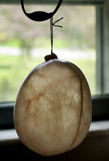
Well apparently it is because originally (for rugby, which later translated to American football) the ball was a leather-covered pig's bladder. 

inflated pig bladder (source)

As you can see, the pig bladder is sort of ovoid in shape.
Further refinements on the shape, like the pointed ends and the elongated shape came afterwards, but the oval rather than spherical nature of the original ball is likely based in pig biology. 

This rugby and football related information was from here and here.

Thursday, February 2, 2012

You can't trust your receptors: Smell

Food smells better when you're hungry, right? This is a common phenomenon that everyone I've ever talked to on the subject has experienced. For a long time, I assumed that the entire process underlying this phenomenon is in the brain proper, and not in the olfactory epithelium (that is, the smell receptors themselves).  However, a study on the adorable (and totally weird) salamander known as the 'Axolotl' suggests that the brain proper can actually modulate how sensitive those smell receptors are.
Axolotls (source)
yes, it does make a good pokemon character


Before I start explaining, let it be known that I am not saying the brain proper doesn't contribute to the 'food-smells-better-when-you're-hungry' phenomenon, in fact I would be very surprised if it didn't involve modulation of the ventral tegmental area, nucleus accumbens, and hypothalamus.

Mousley et al. (2006) use a technique called electro-olfactogram (EOG) to record the signals from smell receptors.  When the cells are excited by an odor, the size of the response can be recorded. They are using this technique in Axolotls, but it can be used in humans too:
EOG recording in humans (source)
Using this technique, Mousley et al. tested whether the size of the smell cells' signal could be modulated by a neuropeptide that is found in the terminal nerve (the nerve that connects the brain proper to the smell-sensing cells).  Chemicals that 'act like' this peptide can have confounding side effects, so the experimentors went to a lot of trouble to make sure they were using the peptide that is actually expressed in these animals.  They copied and synthesized this peptide from the genome of the axolotl. 

So what did they find? The found that this peptide (NPY) could modulate the size of the EOG response in hungry axolotls.  They applied the same amount of odor molecule and the same amount of NPY for each recording, so the increase in response is not due to more odor molecules or more NPY being present.  They suggest that it might be due to mory NPY receptors on the smell cells themselves, indicating that when hungry, the  smell cells change in these animals. 


Mousley et al., 2006 Fig4

So what does that mean? It means that when the animal is hungry, the brain proper has the ability to change the excitability of the smell receptors by dropping some NPY on them (through the terminal nerve).

This study showed the one specific peptide had an effect, but the principle that the brain can actually change the way the peripheral receptors sense things really struck me.  I had always thought that the receptors were pretty much stable, and pretty much always sent the same signal to the brain, but that the way the brain interpreted  that signal could be different. It fundamentally changed my view of sensory cells to learn that the smell receptors don't always send the same signals to the brain. 

source
  It made me rethink the studies that show expectation of taste changes the interpretation. The north dakota wine studies, and the wine-taste evaluation studies show that people rate things differently depending on what they are expecting. A common response is 'ha, what idiots to be influenced by the label of the wine and not trust their own tastebuds' when one reads about studies that show people evaluate white wines with red food coloring as if they were red wines and the like.

However, now I give these wine tasters more credit.  Perhaps the untrustworthy smell cells were actually altered by the brain's expectation. I haven't seen any study testing this idea with EOGs on humans, but I think it would make a great experiment.   


This post was chosen as an Editor's Selection for ResearchBlogging.org

 
ResearchBlogging.orgMousley A, Polese G, Marks NJ, & Eisthen HL (2006). Terminal nerve-derived neuropeptide y modulates physiological responses in the olfactory epithelium of hungry axolotls (Ambystoma mexicanum). The Journal of neuroscience : the official journal of the Society for Neuroscience, 26 (29), 7707-17 PMID: 16855098