Due to starting a major simulation this summer, I am not going to the annual society for neuroscience meeting in San Diego this year. And therefore I won't be neuroblogging it like I did last year.
I look forward to reading the posts and tweets from the official neurobloggers this year.
Here they are:
From Brains to Beyonce by @Spork15
House of Mind by @houseofmind
Neuron Physics by @Eric_Melonakos
NeuroscienceDC by @NeuroscienceDC
Neurolore by @TheMrsZam
NeuroCultureBlog by @LaSaks87
Churchland lab by @anne_churchland
Dormivigilia by @beastlyvaulter
Neurorexia by @ShellyFan
On Psychology and Neuroscience by @astroglia
Más Ciencia por México by @mrenteria_
Imagining Science by @DrImmySmith
Corona Radiata by @JohnKubie
Follow the #SfN13 hashtag on twitter to find all the unofficial coverage of the conference.
© TheCellularScale

Showing posts with label neuroscience for everyone. Show all posts
Showing posts with label neuroscience for everyone. Show all posts
Thursday, November 7, 2013
Monday, May 27, 2013
What is an Experiment?
What is an experiment?
People use the term 'experiment' to meant a lot of things. One may say "she experimented with drugs" or "she performs experimental music". Someone might 'experiment with ones hair' or 'perform a thought experiment'.
All of these uses of the word experiment have distinct connotations, but most of them essentially mean 'to try something and see what happens'. In the examples above, most of the phrases also imply that the experiment is something new. If she experiments with her hair, she's probably trying some new style and seeing if she likes it. If she performs experimental music, she's probably not following the conventional rules for the music she is playing.
Are these kinds of 'experiments' different from the experiments that scientists do? Well yes and no. The basic definition of an experiment as 'trying something [often new] and seeing what happens' is pretty much what scientists do. So what's different? Why isn't someones 'hair experiment' publishable in a scientific journal?
Mythbusters would have you believe that the only difference between science and screwing around is writing it down:
And that is sort of true.
But what really makes a scientific experiment scientific is controls. In our hair example, you can experiment with your hair by dying it black, and seeing if you like it. But that's not the scientific experiment. To be scientific you would have to decide how to measure how much you like your new hair color. You could do this by filling out a survey each day asking you how many times you thought you were pretty or rating your confidence on a scale of 1-10. You could fill this survey out for a week and then dye your hair and fill the survey out for another week. You could then compare the scores and decide if the new black hair had a 'significant' impact on your self-image.
Lets say it does impact your self image and you report higher self-confidence that second week. But what if you feel different just because you have a new hair color, not because you have black hair?
Well, you would want to do a control experiment, which controls for the newness of the hair color. You could control for novelty by dying your hair yet a different color, and taking the survey for another week. Or you could take the survey two months after you dyed your hair black to see if you still report higher confidence or if your confidence has dropped back down to normal.
This is not a perfect experiment by any means, it's not even a clever or well-designed one, but it is somewhat scientific. And illustrates what I think is the most important difference between experimenting as in trying something new, and experimenting as in trying to find something out:
The control group
In addition, here is a great example of how important the control group is in science. (See the epilogue)
© TheCellularScale
 |
| Experimenting with color (source) |
People use the term 'experiment' to meant a lot of things. One may say "she experimented with drugs" or "she performs experimental music". Someone might 'experiment with ones hair' or 'perform a thought experiment'.
All of these uses of the word experiment have distinct connotations, but most of them essentially mean 'to try something and see what happens'. In the examples above, most of the phrases also imply that the experiment is something new. If she experiments with her hair, she's probably trying some new style and seeing if she likes it. If she performs experimental music, she's probably not following the conventional rules for the music she is playing.
Are these kinds of 'experiments' different from the experiments that scientists do? Well yes and no. The basic definition of an experiment as 'trying something [often new] and seeing what happens' is pretty much what scientists do. So what's different? Why isn't someones 'hair experiment' publishable in a scientific journal?
Mythbusters would have you believe that the only difference between science and screwing around is writing it down:
And that is sort of true.
But what really makes a scientific experiment scientific is controls. In our hair example, you can experiment with your hair by dying it black, and seeing if you like it. But that's not the scientific experiment. To be scientific you would have to decide how to measure how much you like your new hair color. You could do this by filling out a survey each day asking you how many times you thought you were pretty or rating your confidence on a scale of 1-10. You could fill this survey out for a week and then dye your hair and fill the survey out for another week. You could then compare the scores and decide if the new black hair had a 'significant' impact on your self-image.
Lets say it does impact your self image and you report higher self-confidence that second week. But what if you feel different just because you have a new hair color, not because you have black hair?
Well, you would want to do a control experiment, which controls for the newness of the hair color. You could control for novelty by dying your hair yet a different color, and taking the survey for another week. Or you could take the survey two months after you dyed your hair black to see if you still report higher confidence or if your confidence has dropped back down to normal.
This is not a perfect experiment by any means, it's not even a clever or well-designed one, but it is somewhat scientific. And illustrates what I think is the most important difference between experimenting as in trying something new, and experimenting as in trying to find something out:
The control group
In addition, here is a great example of how important the control group is in science. (See the epilogue)
© TheCellularScale
Saturday, April 27, 2013
LMAYQ: Mirror Neurons
Mirror neurons really excite people. They've been hyped as the root of empathy and essential to human nature. I've addressed some of this hype, but questions remain. So for this edition of Let Me Answer Your Questions, we will focus on mirror neurons. As always, the LMAYQ series can be found here.
1. "What do mirror neurons look like?"
Good question, and guess what? I have addressed this directly.
2. "Do mirror neurons fire when you die?"
Another good question. Ultimately, all neurons stop firing when you die including mirror neurons. But this doesn't happen immediately. In fact, if the death is due to something traumatic such as decapitation, the neurons might fire more when the nerves are severed between the spinal cord and the brain. But this just brings up questions about the moment of death. Is it when the heart stops, the head is severed? or is it when the neurons stop firing? Can a 'person' be dead when some of their cells are still alive?
In a lot of cellular-level research, cells are kept alive after the animal that they came from has died. Electrophysiologists keep slices of brain alive for hours to record electrical signals from their neurons. Still other projects involve culturing neurons that have been extracted from an animal. These neurons are carefully tended for days, weeks, and even months. These neurons not only stay alive in little dishes, but they can also grow and even control robots.
3. "what does it mean to have a mirrored brain?"
Well. nothing really. I have never heard the term 'mirrored brain' before, and it sounds like something that might be in a pseudo-scientific quiz along the lines of Are you left brained or right brained? "Do you have a mirrored brain? take our quiz and find out"
4. "Is love nothing but mirror cells?"
I love and hate these kinds of questions. The idea that love is nothing if it can be explained by a biological mechanism really gets me. If love is just neurons firing (mirror or otherwise), so what? Why would that make LOVE any less meaningful?
On the other hand, this is a really interesting question if it is asking whether mirror neurons have anything to do with love. Again mirror neurons are neurons that fire when you do something and also when you see someone else do that thing. Specifically, they were discovered in monkeys when monkeys reached for something and then saw other hands reach for something. Then the concept got hyped up. It's easy to imagine that if you have neurons that fire when you do something and when you see some one else do that same thing, that those might have something to do with 'feeling another's pain' and thus empathy. So it's not a huge step to take from there to think that maybe mirror neurons could have something to do with building relationships and love.
But the speculation here is WAY beyond the science. There isn't good solid evidence for mirror neurons controlling empathy, and certainly not for being the basis of love.
© TheCellularScale
 |
| Escher's Mirror (source) |
1. "What do mirror neurons look like?"
Good question, and guess what? I have addressed this directly.
2. "Do mirror neurons fire when you die?"
Another good question. Ultimately, all neurons stop firing when you die including mirror neurons. But this doesn't happen immediately. In fact, if the death is due to something traumatic such as decapitation, the neurons might fire more when the nerves are severed between the spinal cord and the brain. But this just brings up questions about the moment of death. Is it when the heart stops, the head is severed? or is it when the neurons stop firing? Can a 'person' be dead when some of their cells are still alive?
In a lot of cellular-level research, cells are kept alive after the animal that they came from has died. Electrophysiologists keep slices of brain alive for hours to record electrical signals from their neurons. Still other projects involve culturing neurons that have been extracted from an animal. These neurons are carefully tended for days, weeks, and even months. These neurons not only stay alive in little dishes, but they can also grow and even control robots.
 |
| There are living neurons in there (source) |
Well. nothing really. I have never heard the term 'mirrored brain' before, and it sounds like something that might be in a pseudo-scientific quiz along the lines of Are you left brained or right brained? "Do you have a mirrored brain? take our quiz and find out"
4. "Is love nothing but mirror cells?"
I love and hate these kinds of questions. The idea that love is nothing if it can be explained by a biological mechanism really gets me. If love is just neurons firing (mirror or otherwise), so what? Why would that make LOVE any less meaningful?
 |
| Heart Mirror (source) |
But the speculation here is WAY beyond the science. There isn't good solid evidence for mirror neurons controlling empathy, and certainly not for being the basis of love.
© TheCellularScale
Friday, March 15, 2013
Is it 'Important' or is it 'valuable'?
We've recently discussed dopamine as a reward prediction signal. But that is really just the start of the complicated dopamine story.
Some research groups have also found that dopamine neurons respond to aversive stimuli, like an air puff to the face or an electric shock. This finding seems to be be completely incompatible with the idea that dopamine is a signal for reward.
Luckily some scientists took the time to try to resolve this discrepancy. Bromberg-Martin, Matsumoto, and Hikosaka (2010) have written an excellent review paper explaining that some dopamine neurons do code for value (reward), but other dopamine neurons code for salience (importance).
When researchers are recording from a value coding dopamine neuron, it looks like the neuron responds to reward and actually reduces its response to the air puff. This makes sense as a 'dopamine = good' signal.
However, when a researcher is recording from a salience coding dopamine neuron, it looks like the neuron is responding equally to the good thing (reward) and the bad thing (air puff). This is confusing if you think 'dopamine = good', but makes sense if you think 'dopamine = important'. When the cue comes on (a light or a tone that signifies a reward is coming next or an air puff is coming next), these dopamine neurons fire if that cue means something.
Instead of just being confused about why sometimes dopamine would code for value and sometimes it would code for salience, Hikosaka's group showed that these two types of neurons are actually separate populations, and even seem separated in space.
The value dopamine neurons are more ventral in the (monkey) brain, while the salience dopamine neurons are more dorsal-lateral. Importantly these two populations of neurons go to slightly different parts of the striatum and receive signals from different parts of the brain. The review paper suggests that the salience coding neurons receive their input from the central nucleus of the amygdala, while the value coding neurons receive their input from the lateral habenula-RMTg pathway.
The important thing here is that dopamine does not do just one thing to the brain. It doesn't just tell the rest of the brain 'yay, you won!' or 'you want that' etc... It says different things depending on different specific conditions.
Dopamine doesn't 'mean' anything, the cell it comes from and the cell it goes to are what determine what it does. It certainly can't be classified as the 'love molecule'
© TheCellularScale

Bromberg-Martin ES, Matsumoto M, & Hikosaka O (2010). Dopamine in motivational control: rewarding, aversive, and alerting. Neuron, 68 (5), 815-34 PMID: 21144997
 |
| Dopamine's role in reward and punishment (by the hiking artist) |
Luckily some scientists took the time to try to resolve this discrepancy. Bromberg-Martin, Matsumoto, and Hikosaka (2010) have written an excellent review paper explaining that some dopamine neurons do code for value (reward), but other dopamine neurons code for salience (importance).
 |
| Differential Dopamine Coding (Bromberg-Martin et al., 2010 Fig 4) |
When researchers are recording from a value coding dopamine neuron, it looks like the neuron responds to reward and actually reduces its response to the air puff. This makes sense as a 'dopamine = good' signal.
However, when a researcher is recording from a salience coding dopamine neuron, it looks like the neuron is responding equally to the good thing (reward) and the bad thing (air puff). This is confusing if you think 'dopamine = good', but makes sense if you think 'dopamine = important'. When the cue comes on (a light or a tone that signifies a reward is coming next or an air puff is coming next), these dopamine neurons fire if that cue means something.
Instead of just being confused about why sometimes dopamine would code for value and sometimes it would code for salience, Hikosaka's group showed that these two types of neurons are actually separate populations, and even seem separated in space.
 |
| (Bromberg-Martin et al., 2010 Fig 7B) |
The important thing here is that dopamine does not do just one thing to the brain. It doesn't just tell the rest of the brain 'yay, you won!' or 'you want that' etc... It says different things depending on different specific conditions.
Dopamine doesn't 'mean' anything, the cell it comes from and the cell it goes to are what determine what it does. It certainly can't be classified as the 'love molecule'
© TheCellularScale
Bromberg-Martin ES, Matsumoto M, & Hikosaka O (2010). Dopamine in motivational control: rewarding, aversive, and alerting. Neuron, 68 (5), 815-34 PMID: 21144997
Wednesday, February 27, 2013
GABA, how exciting!
I would like to thank my good friend Anonymous for asking me a great question on a previous post.
Anonymous asks:
While I don't know of any instances of glutamate (excitatory) activating GABA (inhibitory) receptors or of GABA activating glutamate receptors, there is an interesting little way that GABA can activate an inhibitory receptor, but actually help excite the cell.
Here's how that works: GABA(A) receptors are permeable to chloride ions, and as the picture above shows, chloride ions (Cl-) are negatively charged. When GABA binds to the receptor, the receptor opens and chloride ions rush in, bringing their negative charge with them. This hyperpolarizes the cell, meaning it brings it lower and lower in total charge (membrane potential), which brings it further and further away from the threshold where it will fire an action potential.
BUT.... if there is a lot of chloride inside the cell already (or if the cell is resting more negatively than the chloride reversal potential), chloride will actually flow out of the cell, bringing its negative charge with it. Negative ions flowing out of the cell will depolarize the neuron increasing its total charge (membrane potential), which brings it closer and closer to the threshold where it will fire an action potential.
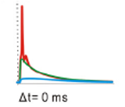
A paper published last year in the Journal of Neuroscience shows that in a model of a hippocampal neuron, when a strong excitatory (glutamate) stimulation happens right after a GABA stimulation close by on the dendrite, the cell is actually more likely to fire than when the glutamate stimulation occurs on its own. This effect is dependent on the location of the GABA stimulation along the dendrite.
This figure shows that a GABA stimuation (first dotted line, blue trace) can push the glutamate (excitatory) stimulation (second dotted line, red trace) up to the point of firing an action potential (green trace). This paper also showed that GABA can still inhibit the action potential in these cells, it just has to be at the soma and almost the same time as the glutamatergic input.
So there you have it, GABA enhancing the likelihood of an action potential and acting excitatory sometimes, and acting inhibitory other times.
© TheCellularScale
Chiang PH, Wu PY, Kuo TW, Liu YC, Chan CF, Chien TC, Cheng JK, Huang YY, Chiu CD, & Lien CC (2012). GABA is depolarizing in hippocampal dentate granule cells of the adolescent and adult rats. The Journal of neuroscience : the official journal of the Society for Neuroscience, 32 (1), 62-7 PMID: 22219270
Anonymous asks:
"Are there any known transmitters in the NS that activate both inhibitory receptor subtypes AND excitatory receptor subtypes? Or does every known transmitter activate EITHER a bunch of excitatory subtypes OR a bunch of inhibitory subtypes?"(btw. This doesn't qualify as a LMAYQ post because it's a real true question that someone directly asked, not a search term)
While I don't know of any instances of glutamate (excitatory) activating GABA (inhibitory) receptors or of GABA activating glutamate receptors, there is an interesting little way that GABA can activate an inhibitory receptor, but actually help excite the cell.
 |
| GABA receptor (source) |
Here's how that works: GABA(A) receptors are permeable to chloride ions, and as the picture above shows, chloride ions (Cl-) are negatively charged. When GABA binds to the receptor, the receptor opens and chloride ions rush in, bringing their negative charge with them. This hyperpolarizes the cell, meaning it brings it lower and lower in total charge (membrane potential), which brings it further and further away from the threshold where it will fire an action potential.
BUT.... if there is a lot of chloride inside the cell already (or if the cell is resting more negatively than the chloride reversal potential), chloride will actually flow out of the cell, bringing its negative charge with it. Negative ions flowing out of the cell will depolarize the neuron increasing its total charge (membrane potential), which brings it closer and closer to the threshold where it will fire an action potential.
 |
| GABA reversing at -62mV (source) |
A paper published last year in the Journal of Neuroscience shows that in a model of a hippocampal neuron, when a strong excitatory (glutamate) stimulation happens right after a GABA stimulation close by on the dendrite, the cell is actually more likely to fire than when the glutamate stimulation occurs on its own. This effect is dependent on the location of the GABA stimulation along the dendrite.
 |
| Chiang et al., 2012 Figure 4E (GPSP in the dendrite) |
This figure shows that a GABA stimuation (first dotted line, blue trace) can push the glutamate (excitatory) stimulation (second dotted line, red trace) up to the point of firing an action potential (green trace). This paper also showed that GABA can still inhibit the action potential in these cells, it just has to be at the soma and almost the same time as the glutamatergic input.
 |
| Chiang et al., 2012 Figure 4G (GPSP in the soma) |
So there you have it, GABA enhancing the likelihood of an action potential and acting excitatory sometimes, and acting inhibitory other times.
© TheCellularScale
Saturday, February 2, 2013
LMAYQ: Let me do your homework for you
Sometimes reading the textbook is just too hard. And sometimes it's much easier just to type your exact homework question into a search engine and find the answer. Before we get started you might want to take a look at Smith and Wren (2010) "What is Plagiarism and how can I avoid it?"
This edition of Let Me Answer Your Questions will address 'homework questions.' As always, you can find previous LMAYQ questions here.
This edition of Let Me Answer Your Questions will address 'homework questions.' As always, you can find previous LMAYQ questions here.
 |
| Tough Homework Questions are for the Internet (source) |
| 1. "cells that fire together a) wire together. b) definitely don’t wire together. c) become overheated and die. d) wire with inactive cells." Ok, student. If you have to turn to the Internet for this multiple choice question, you need a serious lesson in test-taking skills. Here's a tip. If you don't know the answer, eliminate some because they are obviously not the right answer. A good rule to follow is if it says definitely or all or always in the answer it is unlikely that that is the right one. So we can eliminate B. If you know anything about neurons from your class, you should at least know that neurons fire. If they didn't fire they wouldn't 'work'. So you can eliminate C. If all the cells that fired together overheated and died, you would basically not have any neurons left in your brain after reading this sentence. Now you have a 50-50 chance of guessing it correctly. Not too bad. But seriously, if one answer creates a cute little rhyme with the question... it's probably going to be that one. So yes, neurons that fire together wire together. 2. "explain simply where the hippocampus is" The hippocampus is one of the most famous brain structures because it has to do with encoding new memories. It's name comes from the Greek hippo (horse) and campus (sea), because it looks like a seahorse:
3. "Why are neurons and blood cells structured and shaped differently from each other?"
Well basically neurons need to receive and transfer information, while blood cells need to physically move to transfer oxygen. Neurons stay in place, while blood cells travel all over the body. Blood cells need to be small and hydrodynamic to float through your blood vessels. Neurons need to 'cover space' to contact many other neurons, so they have branching dendrites and axons. © TheCellularScale |


Monday, January 7, 2013
Does a high fat diet lead to a less 'rewarding' life?
Some interesting research out of the University of Pennsylvania suggests that a high fat diet can disrupt dopamine signalling.
As I briefly discussed during my SfN Neuroblogging binge, a high fat diet can alter dopamine levels in the brain. To expand on this, we'll look at new research on how exactly this might happen and which specific areas of the brain are affected.
Vucetic et. al. (2012) tested the levels of dopamine-related gene expression (via mRNA) in the hypothalamus and the ventral tegmental area (VTA). The hypothalamus is important because it controls your levels of hunger as well as many other things. The VTA is important because it is the main source of dopamine to the ventral striatum (AKA the Nucleus Accumbens). The VTA-nucleus accumbens pathway is generally thought to signify 'reward' when it is activated. Sex, Drugs, Music, and lots of other 'pleasurable' activities all activate this pathway. So alterations in the dopamine levels here might change how 'rewarded' a person (or mouse in this case) feels in response to pleasurable stimuli.
So Vucetic et al., (2012) found that in the VTA, the levels of tyrosine hydroxylase ("TH", an enzyme indicative of how much dopamine can be made) and dopamine active transporter ("DAT", which gets rid of excess dopamine at the synapse) are both reduced in the mice eating the high fat diet.
By contrast, in the hypothalamus, TH and DAT are both increased due to the high fat diet.
So what does this mean? The authors point out that increased dopamine in the hypothalamus actually promotes eating. Consistent with this idea, the authors show that mice eating the high fat diet actually ate more frequently and ate more total food. Secondly, when there is less dopamine in the VTA, it is likely that a rewarding stimuli will seem less rewarding.
In the author's words:
It is truly a vicious cycle.
*Note: They also look at epigenetic effects on the TH and DAT promoter DNA. If you are interested in that aspect of the study, comment and I can do a follow-up post explaining it, or you can just read the study for yourself, following the link below.
© TheCellularScale
Vucetic Z, Carlin JL, Totoki K, & Reyes TM (2012). Epigenetic dysregulation of the dopamine system in diet-induced obesity. Journal of neurochemistry, 120 (6), 891-8 PMID: 22220805
 |
| This high-fat fed rat sure looks happy to me (source) |
Vucetic et. al. (2012) tested the levels of dopamine-related gene expression (via mRNA) in the hypothalamus and the ventral tegmental area (VTA). The hypothalamus is important because it controls your levels of hunger as well as many other things. The VTA is important because it is the main source of dopamine to the ventral striatum (AKA the Nucleus Accumbens). The VTA-nucleus accumbens pathway is generally thought to signify 'reward' when it is activated. Sex, Drugs, Music, and lots of other 'pleasurable' activities all activate this pathway. So alterations in the dopamine levels here might change how 'rewarded' a person (or mouse in this case) feels in response to pleasurable stimuli.
So Vucetic et al., (2012) found that in the VTA, the levels of tyrosine hydroxylase ("TH", an enzyme indicative of how much dopamine can be made) and dopamine active transporter ("DAT", which gets rid of excess dopamine at the synapse) are both reduced in the mice eating the high fat diet.
 |
| Vucetic et al. (2012) Figure 1 |
So what does this mean? The authors point out that increased dopamine in the hypothalamus actually promotes eating. Consistent with this idea, the authors show that mice eating the high fat diet actually ate more frequently and ate more total food. Secondly, when there is less dopamine in the VTA, it is likely that a rewarding stimuli will seem less rewarding.
In the author's words:
"Collectively, these behaviors have the potential to promote obesity in two distinct ways: (i) through an increase in food intake and (ii) by increasing the drive for palatable food, as the animal with a blunted response to palatable foods may seek and/or consume these food relatively more than a normal animal in order to reach the same rewarding response. "So basically the mice aren't obese because the food they are eating is high fat, they are obese because they are eating MORE food. But of course, they are eating more food because the high fat diet makes them 'want' to eat more food, so the high fat diet is indirectly causing the weight gain.
It is truly a vicious cycle.
*Note: They also look at epigenetic effects on the TH and DAT promoter DNA. If you are interested in that aspect of the study, comment and I can do a follow-up post explaining it, or you can just read the study for yourself, following the link below.
© TheCellularScale
Vucetic Z, Carlin JL, Totoki K, & Reyes TM (2012). Epigenetic dysregulation of the dopamine system in diet-induced obesity. Journal of neurochemistry, 120 (6), 891-8 PMID: 22220805
Thursday, December 13, 2012
LMAYQ: seriously deep questions
And now, let me answer your Seriously Deep Questions. All questions answered can be found in the LMAYQ index. And as always these are real true search terms that the all-knowing Internet directed to The Cellular Scale. Let's begin.
1. "Do thoughts look like trees?"
Great question. Lots of things look like trees, certainly neurons do. But thoughts themselves?
It is my personal opinion that thoughts do not actually look like anything. I've dissected many a brain and haven't ever seen one. However, let's suppose thoughts look like something, what would they look like?
One possibility is that the thought looks like what you are thinking about. A pretty ancient idea is that there are actually two of every object, one that is external (the actual object), and one that is internal which is our representation of that object. This can be taken quite literally in which case if you are looking at or thinking about a tree, your thought will look like a tree, but if you are thinking about a dog, your thought will look like a dog. This strikes me as unlikely.
So another way to look at it is what does the brain look like when it is having a thought? In this case there is some support for the 'thought looks like what you are thinking' hypothesis, but it is very limited.
Above is a famous example of how a visual stimulus can be reflected in the brain in a very literal way. In this case a monkey looks at a grid and the activation pattern in the brain looks like a grid. But these days 'thoughts' usually look like this:
And there is no obvious or literal relationship between the shape of the fMRI image and the thought that is thunk.
2. "Why Neuroscience?"
Because neuroscience is our best chance at answering important questions like 'what do thoughts look like?' and 'How do we know what we know?'
3. "Do neurons tell you how to move or do they fire in response?"
Another excellent and deep question. The answer is (of course) that they do both.
 People used to think of the brain as a black box, where sensory input comes in (like through your eyes) and gets 'processed' by the brain and a motor output comes out (like through your hands).
All of these steps, the sensory input, the motor output, and the processing in between take neurons.
But of course there is the Venus flytrap which doesn't have 'neurons' per se, but does receive sensory input and generate motor output.
But the processing part of this process, the black box, is really complicated. There really is an unanswered question there about whether neurons are responding to something or telling something. When studies find that mirror neurons fire 'in response to' seeing actions performed, or that some amygdala neurons fire in response to pictures of animals, the question is always why are these neurons firing? Are the neurons telling another part of the brain 'this is an animal'? or are the neurons responding to that information?
© TheCellularScale
 |
| Thoughts on grass (source) |
Great question. Lots of things look like trees, certainly neurons do. But thoughts themselves?
It is my personal opinion that thoughts do not actually look like anything. I've dissected many a brain and haven't ever seen one. However, let's suppose thoughts look like something, what would they look like?
One possibility is that the thought looks like what you are thinking about. A pretty ancient idea is that there are actually two of every object, one that is external (the actual object), and one that is internal which is our representation of that object. This can be taken quite literally in which case if you are looking at or thinking about a tree, your thought will look like a tree, but if you are thinking about a dog, your thought will look like a dog. This strikes me as unlikely.
So another way to look at it is what does the brain look like when it is having a thought? In this case there is some support for the 'thought looks like what you are thinking' hypothesis, but it is very limited.
 |
| Do thoughts look like nets? (source) |
 |
| thinking (source) |
2. "Why Neuroscience?"
Because neuroscience is our best chance at answering important questions like 'what do thoughts look like?' and 'How do we know what we know?'
3. "Do neurons tell you how to move or do they fire in response?"
Another excellent and deep question. The answer is (of course) that they do both.

All of these steps, the sensory input, the motor output, and the processing in between take neurons.
But of course there is the Venus flytrap which doesn't have 'neurons' per se, but does receive sensory input and generate motor output.
But the processing part of this process, the black box, is really complicated. There really is an unanswered question there about whether neurons are responding to something or telling something. When studies find that mirror neurons fire 'in response to' seeing actions performed, or that some amygdala neurons fire in response to pictures of animals, the question is always why are these neurons firing? Are the neurons telling another part of the brain 'this is an animal'? or are the neurons responding to that information?
© TheCellularScale
Tuesday, November 20, 2012
Virtual reality for your robot cockroach
I have previously covered some interesting advances in the world of cyborg insects.
Latif and Bozkurt from North Carolina state university recently presented a paper (though I can't find a peer-reviewed publication on Pubmed), explaining their Biobot. They use the Madacascar hissing cockroach...
... and attach a electrically stimulating 'backpack' (see first picture). They then stimulate the the antennae in a variety of ways to 'steer' the Biobot.
Pretty cool. The authors note that generally the cockroaches want to walk straight until they encounter an obstacle (or stimulation). So, sure, this is sort of like steering a horse with reins, but the horse has to be trained to know what the bridle signals mean. This setup is more like creating a virtual reality for the cockroach, where it thinks that it has 'run into' something at certain points on the line. This is similar to creating a virtual reality for worms by stimulating specific neurons with light.
Of course the practical applications of this are a little iffy. People always seems to say that these little insect-bots could be of use in disaster settings where people need to get some ground level surveillance of a rubble-littered area, but I think the scientific applications for this are what is really exciting. Being able to create a virtual reality of any shape or size could allow for tests of spatial navigation in the cockroach. You could even try to train the cockroach to find something or avoid something and the 'confuse it' by changing the virtual environment suddenly. Could it adapt?
© TheCellularScale
 |
| Biobot backpack (cockroach size) (source) |
 |
| Hissing Cockroach (source). Terrifying. |
This is very similar to the backyard brains Roboroach, but the system created by Latif and Bozkurt is extremely precise. Rather than just making the Biobot turn when stimulated, Latif and Bozkurt can make the cockroach walk a specified line.
"In these studies, electrical pulses were applied to the insect to create biomechanical or sensory perturbations in the locomotory control system to steer it in desired directions, similar to steering a horse with bridle and reins." -Latif and Bozkurt
Pretty cool. The authors note that generally the cockroaches want to walk straight until they encounter an obstacle (or stimulation). So, sure, this is sort of like steering a horse with reins, but the horse has to be trained to know what the bridle signals mean. This setup is more like creating a virtual reality for the cockroach, where it thinks that it has 'run into' something at certain points on the line. This is similar to creating a virtual reality for worms by stimulating specific neurons with light.
Of course the practical applications of this are a little iffy. People always seems to say that these little insect-bots could be of use in disaster settings where people need to get some ground level surveillance of a rubble-littered area, but I think the scientific applications for this are what is really exciting. Being able to create a virtual reality of any shape or size could allow for tests of spatial navigation in the cockroach. You could even try to train the cockroach to find something or avoid something and the 'confuse it' by changing the virtual environment suddenly. Could it adapt?
© TheCellularScale
Thursday, November 8, 2012
why you shouldn't wash your brain with soap
I can't think of any situation where you might be inclined to soap up your brain (except maybe if you had recently been trepanned), but it is still a bad idea.
When used on say, an oily frying pan, soap + scrubbing will trap the oil in little units which can be rinsed off. Without soap, using only water, the oil which is hydrophobic (meaning it would rather stick to anything besides water) will stick to the pan rather than the water.
How does this relate to the brain? Well the cell membrane which helps give the shape to the neurons is made up of a lipid bilayer. These lipids have a hydrophobic tail (which hides in the middle of the layer) and a hydrophilic head which faces outward, just like the oil particles above.
So basically if you scrubbed your brain cells with soap, the membrane that holds the neuron together would be disrupted. Scientists actually use this principle to get stuff (like DNA) out of a neuron. In DNA extraction, there is a lysis step in which a detergent (like SDS) is applied to the tissue and given a good shake. This disrupts the membrane and allows access to the contents of the neuron.
You can wash your skin with soap because the living skin cells are protected by an outer dead skin cell layer. Though if you soap up too much, you can actually dry out yours skin by stripping it of lipids faster that they can be replenished. See "How much should you shower" for an excuse to stay in bed tomorrow morning rather than get up and shower.
© TheCellularScale
 |
| you can actually buy soap shaped like a brain here. (It smells like bubble gum!) |
 |
| Soap + shaking = trapped oil (source) |
 |
| Cell membrane (source) |
You can wash your skin with soap because the living skin cells are protected by an outer dead skin cell layer. Though if you soap up too much, you can actually dry out yours skin by stripping it of lipids faster that they can be replenished. See "How much should you shower" for an excuse to stay in bed tomorrow morning rather than get up and shower.
© TheCellularScale
Friday, October 5, 2012
SfN Neuroblogging 2012: Introduction
| SfN |
What is the Society for Neuroscience meeting?
The Society for Neuroscience annual meeting is the biggest neuroscience conference ever. It is attended by about 30,000 people each year, and there are about 20,000 presentations (including posters). It's like a gigantic science fair.
 |
| one tiny corner of SfN |
I am planning to post at least once a day about interesting posters or talks I see or events I attend during the actual meeting (October 13th-17th). These posts won't be super in-depth in terms of the science, but I plan to write a whole bunch of follow up posts after the meeting delving deeper into the most interesting science.
What will I be blogging about?
SfN is divided up into themes. I don't know about you, but I have never put much thought into the 'theme' I wanted to visit during the meeting. In fact I am not quite certain which theme my own poster is in. But regardless, the official bloggers are also organized by theme and The Cellular Scale will be covering themes B and D.
Theme B: Neural Excitability, Synapses, and Glia: Cellular Mechanisms.
This is a good match for this blog because I've already discussed many of the sub-topics in this theme, such as synaptic plasticity, glia-neuronal interactions, and other weird things about cells.
Theme D: Sensory and Motor Systems
This is also a good match because I love to blog about the senses at a cellular level. See these links for posts about the cells involved in vision, hearing, taste and smell.
So these themes are what I am 'officially' covering, but I plan to blog about the most interesting science even if it falls under another category.
What else?
I am going to index all the "SfN Neuroblogging" posts in a tab at the top of the page so you can easily get to all of them.
I will also be tweeting the meeting as they say, so follow me @cellularscale on twitter for up-to-the-minute updates.
If you have any questions that I didn't answer here, add a comment and I will try to answer it.
© TheCellularScale
Wednesday, September 26, 2012
you can't trust your brain: memory
"Flashbulb" memories are those vivid memories of specific salient events. The 'everyone remembers exactly where they were when...' sort of events. In the USA, and depending on how old you are, you might remember the assassination of JFK, or Martin Luther King Jr. in this way. In this century, most Americans remember exactly where they were when they heard about the 9/11 attacks on the world trade center and pentagon.
It's widely acknowledged these days that the brain is not really a safe place to store information. Memories of events change over time. But for a while the "flashbulb" memory was thought to be immune from the memory-altering properties of time. Think about your own memories of 9/11 or another highly meaningful event. I bet you are pretty certain about the details. I, for example, was in my second year of college and I know exactly who told me that the first tower was hit, exactly where I was standing on the quad, and exactly what class I was going to....
...or do I?
A study in 2003 tested the consistency of flashbulb memories over time and compared the details to 'control memories' of everyday events. They specifically recorded memories from people during the day after the 9/11 attacks, and then recorded memories of the same events from subsets of those same people 1 week, 6 weeks, and 32 weeks later. They found that the flashbulb memories did have different properties when compared to control memories, but that consistency was not one of them.
Talarico and Rubin show that the flashbulb memories and the everyday memories had the same time-dependent decay (that x axis is in days), demonstrating that the flashbulb memory did not have some special property that protected it from corruption.
However, they did find that the level of confidence in the memory was higher for flashbulb memories than for everyday memories. People thought (incorrectly) that their memories of the 9/11 attacks were more accurate than their other memories.
So again we learn the lesson that we cannot trust ourselves.
In the authors words:
But I think the most interesting finding in this paper was that the flashbulb memories of 9/11 were more likely to be recalled 'through ones own eyes' than the everyday memories. Everyday memories were seen 'through ones own eyes' at the beginning and a at 1 week, but at 6 and 32 weeks the everyday memories were more likely to be seen 'from an outside observer perspective.' The flashbulb memories, on the other hand, were seen 'through ones own eyes' at all time points. Indeed, when I think of my own 9/11 memory, I still see it through my own eyes.
The authors don't go into why that might be or what it might mean, so we are left to wonder.
© TheCellularScale
Talarico JM, & Rubin DC (2003). Confidence, not consistency, characterizes flashbulb memories. Psychological science, 14 (5), 455-61 PMID: 12930476
 |
| "Never Forget" (source) |
...or do I?
A study in 2003 tested the consistency of flashbulb memories over time and compared the details to 'control memories' of everyday events. They specifically recorded memories from people during the day after the 9/11 attacks, and then recorded memories of the same events from subsets of those same people 1 week, 6 weeks, and 32 weeks later. They found that the flashbulb memories did have different properties when compared to control memories, but that consistency was not one of them.
 |
| Talarico and Rubin 2003, Figure 1a |
However, they did find that the level of confidence in the memory was higher for flashbulb memories than for everyday memories. People thought (incorrectly) that their memories of the 9/11 attacks were more accurate than their other memories.
So again we learn the lesson that we cannot trust ourselves.
In the authors words:
"The true 'mystery,' then, is not why flashbulb memories are so accurate for so long,... but why people are so confident in the accuracy of their flashbulb memories." Talarico and Rubin (2003)
But I think the most interesting finding in this paper was that the flashbulb memories of 9/11 were more likely to be recalled 'through ones own eyes' than the everyday memories. Everyday memories were seen 'through ones own eyes' at the beginning and a at 1 week, but at 6 and 32 weeks the everyday memories were more likely to be seen 'from an outside observer perspective.' The flashbulb memories, on the other hand, were seen 'through ones own eyes' at all time points. Indeed, when I think of my own 9/11 memory, I still see it through my own eyes.
The authors don't go into why that might be or what it might mean, so we are left to wonder.
© TheCellularScale
Talarico JM, & Rubin DC (2003). Confidence, not consistency, characterizes flashbulb memories. Psychological science, 14 (5), 455-61 PMID: 12930476
Sunday, August 26, 2012
LMAYQ: Safety First
Welcome to part 3 of the million part series "Let Me Answer Your Questions" (LMAYQ). Here I address questions that people have asked The Internet. The Internet, in turn, directed these people to The Cellular Scale, where their questions were... not answered.
Today our topic is:

These 3 questions inspire me to warn you readers about a couple dangerous things.
1. "Will Parkinson's doctor let me have DBS?"
This is a really really important question because I absolutely do not know the answer to this. It must have brought someone to my post lauding the amazing power of Deep Brain Stimulation and its ability to stop Parkinson's Disease symptoms in their tracks. This is such an important question because it has prompted me to make this point:
I am not a clinical doctor and more importantly The Internet is not your doctor and you should be careful when you ask The Internet medical questions. Yes DBS has shown amazing results, but its long term side effects are not even known yet and it requires a very serious surgical procedure. Just because I think its an amazing step forward, doesn't mean I think you should get it. I don't know any thing about you or your condition (and neither does anyone else on The Internet).
2. "Is DBS a cure for Parkinson's Disease?"
Following up on question 1, I titled my blog post "How does DBS cure Parkinson' Disease?", and focused on some new research looking into its mechanism of action. It was somewhat careless of me to use the word 'cure' all throughout the post, when really it should have been 'treatment'. The most accurate answer to your question is "DBS treats Parkinson's Disease symptoms"
3. "What happens when you give a mouse cocaine?"
This is a good and interesting question, but also inspires me to make a warning statement:
Today our topic is:
These 3 questions inspire me to warn you readers about a couple dangerous things.
1. "Will Parkinson's doctor let me have DBS?"
This is a really really important question because I absolutely do not know the answer to this. It must have brought someone to my post lauding the amazing power of Deep Brain Stimulation and its ability to stop Parkinson's Disease symptoms in their tracks. This is such an important question because it has prompted me to make this point:
This blog is not giving medical advice.
2. "Is DBS a cure for Parkinson's Disease?"
Following up on question 1, I titled my blog post "How does DBS cure Parkinson' Disease?", and focused on some new research looking into its mechanism of action. It was somewhat careless of me to use the word 'cure' all throughout the post, when really it should have been 'treatment'. The most accurate answer to your question is "DBS treats Parkinson's Disease symptoms"
3. "What happens when you give a mouse cocaine?"
This is a good and interesting question, but also inspires me to make a warning statement:
Do not try this at home.
Do NOT give your pet mice random drugs (even legal ones). It is not healthy for them.
This question brought someone to the post "If you give a mouse a placebo..." which discusses how to trick a mouse into thinking it is getting cocaine. This is really important research isolating the effect of the actual cocaine directly acting on the brain compared to the effect of the inactive cocaine acting indirectly on the brain.
The answer is: If you give a mouse cocaine, it can get addicted to it, which is why mice are used to study the effects of cocaine addiction and the efficacy of possible treatments.
But really I want to use this question as an opportunity to explain how research on animals is different from just giving animals drugs.
All the research I report on here is peer-reviewed. This means other scientists besides the authors have read it carefully, and had an opportunity to point out any flaws or weak points in the research design or execution. In addition, research that uses animals adheres to extensive animal usage and handling regulations. It's not like someone just thinks 'dude, I wonder what would happen if you gave a mouse this or that' and then does and calls it research. Before scientists uses a single animal for any research, they have to write a thorough (usually 6 pages or so) explanation for exactly how many animals they are using and why they have to use that many and what they are going to use them for. They have to list the steps they will take to minimize pain and explain how they will ensure that the animals are treated humanely. Then that document is read and discussed by a whole committee whose job it is to make sure that animals are being treated well at that institute. The committee then can either approve the animal use, or reject it and ask the scientist to only use this many instead of that many or use this kind of surgery instead of that kind of surgery.
So there you have it, some warnings about what you should and shouldn't draw from this blog (and The Internet in general). Stay tuned for more fantastic answers to your fantastic questions.
© TheCellularScale
Sunday, August 12, 2012
How does Deep Brain Stimulation cure Parkinson's Disease? A Cellular Look
Deep Brain Stimulation (DBS) is one of the miracle cures of our lifetime. Used to treat Parkinson's Disease, and now being applied to depression, it is a drastic but amazingly effective measure.
EDIT 9/23/12: Just because DBS has been shown to be amazingly effective in treating Parkinson's Disease symptoms, that does not mean that it is right for everyone. There are other effective treatments for PD and DBS involves an invasive surgery. In addition, the long term effects of having a stimulating electrode constantly pulsing electricity in your brain are not yet fully known. In other words, this blog is not giving medical advice, so please don't take it as such.
To apply DBS, an electrode is implanted deep in the brain (the SubThalamic Nucleus for Parkinson's Disease). An implanted wire under the skin connects the stimulation electrode in the brain to a stimulator implanted on the chest. The stimulation can be turned on and off here.
Here is a short video showing the effect of DBS on and DBS off. (The actual demonstration starts at 1 minute in, the rest is some arguing in French)
Once the stimulation is turned off, the man has twitches an tremors that prevent him from functioning normally. As soon as the stimulation is turned back on, he can walk and talk smoothly.
This is pretty astounding, right? I mean honestly, if I just saw this video and didn't know anything about it, I might not even believe that it was real. How can electrical stimulation in the brain completely alter this person's ability to move?
Well, scientists around the world are wondering the exact same thing. One recent study made use of optogenetics to dissect the pathways involved in deep brain stimulation.
They first elicited Parkinson's Disease in mice by destroying the dopaminergic cells (in the substantia nigra which feeds into the striatum) on only one side of the brain. Then they give the mice a stimulant drug which makes them hyper. While normal mice would run all over the place on this stimulant, these hemi-parkinson's mice run in a circle because of the imbalance between the two sides of the brain.
To test whether the treatment you are giving the mouse 'cures' their Parkinson's Disease, you literally count how many times the mouse runs in a circle. If it runs in a lot of circles, your 'cure' didn't work.
Implanting an electrode into the STN of the mice and applying the stimulation did reduce their circle running, just like the DBS reduces Parkinson's symptoms in humans. But here's the thing, the STN has a lot of neurons in it, and a lot of axons passing through it. Stimulating this brain region could be inhibiting firing, or exciting neurons, or something else entirely.
So Gradinaru et al. (2009) decided to stimulate specific (genetically identified) classes of neurons to determine which aspect of the electrical stimulation was actually 'curing' the Parkinson's symptoms.
First they tested the most common hypothesis, that the stimulation inhibited the STN. However, when they directly inhibited the neurons of the STN, their mice still ran in circles, indicating that the brain was still unbalanced.
Second they stimulated the glial cells around the STN, but still no luck.
Third they excited the neurons of the STN.... but... still the mouse ran in circles.
I imagine this was very puzzling to the scientists conducting this research. If stimulating the STN with a big metal electrode is not exciting, and not inhibiting the STN, what on earth could it be doing?
Finally they tried targeting the axons of the motor cortex which run through the STN. And! low and behold, the mouse did not run in circles any more. Below, rotations per minute is basically a measure of circle running, and the HFS is the stimulation. You can see that as soon as the stimulation is activated the mice generally stopped running in circles, but when the stimulation was turned off, they ran in circles again.
So a new theory has emerged, that the stimulation of the STN might actually be acting on other neurons which are not even located in that brain structure. It clearly works in mice, but whether this is the way that Deep Brain Stimulation works in humans is still not clear.
© TheCellularScale
Gradinaru V, Mogri M, Thompson KR, Henderson JM, & Deisseroth K (2009). Optical deconstruction of parkinsonian neural circuitry. Science (New York, N.Y.), 324 (5925), 354-9 PMID: 19299587
EDIT 9/23/12: Just because DBS has been shown to be amazingly effective in treating Parkinson's Disease symptoms, that does not mean that it is right for everyone. There are other effective treatments for PD and DBS involves an invasive surgery. In addition, the long term effects of having a stimulating electrode constantly pulsing electricity in your brain are not yet fully known. In other words, this blog is not giving medical advice, so please don't take it as such.
 |
| Deep Brain Stimulation (DBS) |
To apply DBS, an electrode is implanted deep in the brain (the SubThalamic Nucleus for Parkinson's Disease). An implanted wire under the skin connects the stimulation electrode in the brain to a stimulator implanted on the chest. The stimulation can be turned on and off here.
Here is a short video showing the effect of DBS on and DBS off. (The actual demonstration starts at 1 minute in, the rest is some arguing in French)
Once the stimulation is turned off, the man has twitches an tremors that prevent him from functioning normally. As soon as the stimulation is turned back on, he can walk and talk smoothly.
This is pretty astounding, right? I mean honestly, if I just saw this video and didn't know anything about it, I might not even believe that it was real. How can electrical stimulation in the brain completely alter this person's ability to move?
Well, scientists around the world are wondering the exact same thing. One recent study made use of optogenetics to dissect the pathways involved in deep brain stimulation.
They first elicited Parkinson's Disease in mice by destroying the dopaminergic cells (in the substantia nigra which feeds into the striatum) on only one side of the brain. Then they give the mice a stimulant drug which makes them hyper. While normal mice would run all over the place on this stimulant, these hemi-parkinson's mice run in a circle because of the imbalance between the two sides of the brain.
 |
| or hemi-parkinson's disease (source) |
To test whether the treatment you are giving the mouse 'cures' their Parkinson's Disease, you literally count how many times the mouse runs in a circle. If it runs in a lot of circles, your 'cure' didn't work.
Implanting an electrode into the STN of the mice and applying the stimulation did reduce their circle running, just like the DBS reduces Parkinson's symptoms in humans. But here's the thing, the STN has a lot of neurons in it, and a lot of axons passing through it. Stimulating this brain region could be inhibiting firing, or exciting neurons, or something else entirely.
So Gradinaru et al. (2009) decided to stimulate specific (genetically identified) classes of neurons to determine which aspect of the electrical stimulation was actually 'curing' the Parkinson's symptoms.
First they tested the most common hypothesis, that the stimulation inhibited the STN. However, when they directly inhibited the neurons of the STN, their mice still ran in circles, indicating that the brain was still unbalanced.
Second they stimulated the glial cells around the STN, but still no luck.
Third they excited the neurons of the STN.... but... still the mouse ran in circles.
I imagine this was very puzzling to the scientists conducting this research. If stimulating the STN with a big metal electrode is not exciting, and not inhibiting the STN, what on earth could it be doing?
Finally they tried targeting the axons of the motor cortex which run through the STN. And! low and behold, the mouse did not run in circles any more. Below, rotations per minute is basically a measure of circle running, and the HFS is the stimulation. You can see that as soon as the stimulation is activated the mice generally stopped running in circles, but when the stimulation was turned off, they ran in circles again.
 |
| Gradinaru et al., 2009 |
So a new theory has emerged, that the stimulation of the STN might actually be acting on other neurons which are not even located in that brain structure. It clearly works in mice, but whether this is the way that Deep Brain Stimulation works in humans is still not clear.
© TheCellularScale
Wednesday, July 25, 2012
Fire Together Wire Together
 |
| Fire together Wire together (source) |
One theory is that neurons strengthen their synapses based on the specific timing between receiving a signal and firing an action potential. This is called Spike Timing Dependent Plasticity (STDP). The basic premise is that if neuron A fires first, and then neuron B fires, neuron A is probably partly responsible for neuron B firing. If this is the case then the signal from A to B probably contains meaningful information.
 |
| Neurons Connecting (modified from here) |
And the opposite? What if neuron B fires before neuron A? It could be a meaningful signal or it could just be random noise. Maybe neuron A fired for no good reason (or even just a little vesicle of glutamate popping out untriggered). Many studies show that when B fires before A, the connection was actually weakened.
So the saying perhaps should be 'neurons that fire one right after the other wire together' ... But that just doesn't flow off the tongue the way 'fire together wire together' does.
STDP is incredibly complex and even though this A before B = strengthening and B before A = weakening explanation makes intuitive sense, there is an exception for every rule. Some studies find just the opposite! And some studies find that both directions strengthen the synapse (A-B and B-A).
Dan and Poo (2006) have a nice table explaining these exceptions and the parts of the brain where they are found.
© TheCellularScale
Bi G, & Poo M (1999). Distributed synaptic modification in neural networks induced by patterned stimulation. Nature, 401 (6755), 792-6 PMID: 10548104
Dan Y, & Poo MM (2006). Spike timing-dependent plasticity: from synapse to perception. Physiological reviews, 86 (3), 1033-48 PMID: 16816145
Sunday, July 22, 2012
Do small men think like big women?
Endless research has been conducted on the neurological differences between women and men. However, a study out of the University of Florida explains that almost all of the anatomical differences previously reported can be accounted for simply by adjusting for total brain size.
A result hardly worth speculating upon.
Leonard et al., 2008 also found some corpus callosum differences between the genders, but when they graphed the size of the corpus callosum against the size of the whole brain...
They found a continuum. The difference in size between the female and male corpus callosum is entirely due to the difference in size of the female and male brain as a whole.
As with Von Economo neurons, maybe brains of different sizes work similarly, but have to be shaped differently to do so.
So rather than wildly speculating that women are better at this or that because they have stronger connections between their hemispheres, we should put our efforts into discovering evolutionary reasons why small men would be better multi-taskers that large men.
© TheCellularScale
DeLacoste-Utamsing C, & Holloway RL (1982). Sexual dimorphism in the human corpus callosum. Science (New York, N.Y.), 216 (4553), 1431-2 PMID: 7089533
Leonard CM, Towler S, Welcome S, Halderman LK, Otto R, Eckert MA, & Chiarello C (2008). Size matters: cerebral volume influences sex differences in neuroanatomy. Cerebral cortex (New York, N.Y. : 1991), 18 (12), 2920-31 PMID: 18440950
 |
| (Lady Gaga is an excellent source of exaggerated imagery) |
Leonard et al., (2008) recruited 100 men and 100 women and imaged their brains. They showed that men generally have larger brains that women (not surprising, men generally have larger bodies than women).
 |
| Leonard et al., 2008 Figure 2 |
But what is fascinating is that when comparing specific regions, the gender of the brain mattered less than the size of the whole brain.
In other words if you had a small male brain, it would look almost indistinguishable from a large female brain. (See their Figure 3)
What I find most interesting in this paper is that it refutes the much purported "Corpus Callosum Myth".
 |
| Corpus Callosum (source) |
The Corpus Callosum is main white matter connection between the two hemispheres of the brain. The "Corpus Callosum Myth" is that female brains have larger corpus callosa than male brains.
I have to admit that I am not immune from gender bias. When I first heard that women had larger corpus callosa than men, my immediate thoughts were towards how that could make sense. I thought "ah, well then maybe that is why women are better at seeing the big picture or at multi-tasking" and other thoughts along those lines.
What I definitely did NOT think was "I bet that was a small, poorly controlled study which did not even reach statistical significance." Well as it turns out, I should have. DeLacoste-Utamsing and Holloway (1982) analyzed only 14 brains (9 male and 5 female), and found that
"The average area of the posterior fifth of the corpus callosum was larger in females than in males (p=0.08)" DeLacoste-Utamsing and Holloway (1982) p. 1431
A result hardly worth speculating upon.
Leonard et al., 2008 also found some corpus callosum differences between the genders, but when they graphed the size of the corpus callosum against the size of the whole brain...
 |
| Figure 3B (female brains white circles, male brains filled squares) |
As with Von Economo neurons, maybe brains of different sizes work similarly, but have to be shaped differently to do so.
So rather than wildly speculating that women are better at this or that because they have stronger connections between their hemispheres, we should put our efforts into discovering evolutionary reasons why small men would be better multi-taskers that large men.
© TheCellularScale
UPDATE (7/23/12): I just want to be perfectly clear. I don't actually think that small men think like women. The whole point of this post is to show that popular studies explaining that 'men and women's brains are different' may sound like they make sense, but there is often another explanation. In this case: if you are going to claim that the size of the corpus callosum means that women are better multi-taskers, then you have to ALSO claim that small men are better multi-taskers. And that large women are worse multi-taskers. (These seem like totally ridiculous claims to me, but feel free to construct an experiment to test these hypotheses).
For more on gender and gender differences (or lack thereof) in the brain, see my previous posts:
Leonard CM, Towler S, Welcome S, Halderman LK, Otto R, Eckert MA, & Chiarello C (2008). Size matters: cerebral volume influences sex differences in neuroanatomy. Cerebral cortex (New York, N.Y. : 1991), 18 (12), 2920-31 PMID: 18440950
Subscribe to:
Comments (Atom)